Christina Gale, BSc, Swine Marketing Manager, Eduardo Velazquez, MRCVS, Swine Veterinary Service Manager and Emma Pattison, BSc, Swine Field and Vaccination Services Manager, Ceva Animal Health, UK
What is porcine enzootic pneumonia and how does it spread in swine herds?
Mycoplasma hyopneumoniae (M. hyopneumoniae) is one of the most important primary pathogens of the porcine respiratory system and is the causative agent of enzootic pneumonia (EP) (Fraile et al., 2010). M. hyopneumoniae is widespread throughout the pig population and is endemic on most farms worldwide. Pneumonic lung lesions due to M. hyopneumoniae are commonly observed in the slaughterhouse, with average herd-level prevalence reported as 24%, ranging up to 88% (Maes et al., 2001), demonstrating the large variability observed between units depending on individual farm situation.
How does M. hyopneumoniae infect the respiratory tract?
Mycoplasma hyopneumoniae is 200–500 nm in size and requires complex media and aerobic and microaerophilic conditions to be cultured. Infection with M. hyopneumoniae occurs via inhalation of infected aerosols or via direct contact with infected animals. The pathogen establishes itself in the respiratory tract, attaching to the ciliated cells of the tracheal, bronchial and bronchiolar epithelium. Once in the respiratory tract, it can persist for weeks to months and causes pathological changes in the lungs. Lesions are thought to form in the lobes of the lung due to lack of normal clearance of secretory products from the respiratory system, due to infection of the ciliated epithelium (Taylor, 1995). Early lesions are observed as small dark red areas in the anterior lobes, which enlarge over time and after a few weeks lose their red colour and become more pink (Figure 1). Lesions resolve after 12 to 14 weeks with formation of interlobular fissures (Maes et al., 2008). This is important to note, as infections early in production may be resolved once the animal reaches the slaughterhouse, so any scarring (Figure 2) should be noted as losses may still have occurred during the growing and finishing stages.
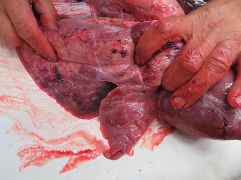
Figure 1 - Pneumonic lung lesions caused by infection with M.hyopneumoniae
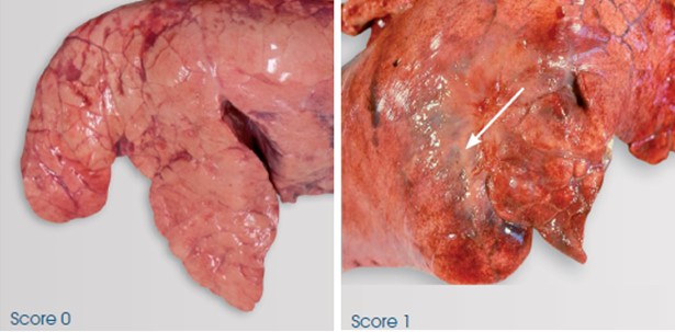
Figure 2 - Scarring of lobes (score 1) due to recovery of lung lesions caused by infection with M.hyopneumoniae
Histologically, inflammation occurs and neutrophils can accumulate in the airways and bronchioles. Lymphocytes and macrophages will also be present 5 days after infection, followed by presence of large mononuclear cells, polymorphs, lymphocytes and plasma cells in the alveoli from day 7 (Taylor, 1995). Immunoglobulin IgA forms in tracheal mucosa so may be detectable from day 30 on diagnostic testing of secretions, and IgG may also be present at a slightly later stage.
What are the clinical signs of enzootic pneumonia in pigs?
Enzootic pneumonia can clinically be acute or chronic in its form, with different clinical presentations of disease:
- Acute form: pigs of all ages can be affected and most likely will experience pyrexia, anorexia and respiratory distress, usually accompanied with a distinct cough (Taylor, 1995).
- Chronic form: few clinical signs may be observed, particularly in young growing pigs, where diarrhoea and a dry cough may be the only visible signs if present. Later in production, this cough may become a barking cough in the finishing house, and what may be most apparent is the variation in size of pen mates, indicating performance has been compromised in some animals.
How is enzootic pneumonia diagnosed?
Diagnosis can be carried out by M. hyopneumoniae-specific seroconversion or laryngeal swabs tested by PCR (Pieters et al., 2017). Examination of the lungs at slaughter can also be useful to report presence of pneumonic lesions, characterised by consolidated areas especially in the cranial lobes (Maes et al., 2001). Seasonal patterns are often observed with M. hyopneumoniae, with lung lesion prevalence and severity increasing following the winter months. This was demonstrated by Ostanello et al. (2007) where, as scored using the Madec and Kobisch method (1982), mean lung lesion score was significantly increased in batches of animals raised in the winter months (October–March) compared to the summer months (April–September), with scores of 2.33 and 1.81 respectively. Decreased growth rate and feed conversion ratio (FCR) may also be observed, typically with no or low mortality (Sibila et al., 2009).
How does enzootic pneumonia trigger co-infections and PRDC in pigs?
Mycoplasma hyopneumoniae is also a major contributor to development of the porcine respiratory disease complex (PRDC) (Garcia-Morante et al., 2015). The pathogen modifies the immune response and facilitates the expression of co-infections. It is described as an inhibitor of macrophage phagocytic activity, which may explain the chronicity of M. hyopneumoniae infections and the greater host susceptibility to other pathogens (Figures 3 and 4) (Saade et al., 2020). Co-infections are very important in respiratory disease, as interaction of multiple pathogens has been shown to increase the severity of clinical signs (Saade et al., 2020) and therefore the impact on performance.
Viral co-infections with M. hyopneumoniae
Co-infections can occur with both viral and bacterial diseases. Regarding viruses, it is important to mention three very important diseases affecting the swine herd globally:
- Porcine Reproductive and Respiratory Syndrome (PRRS): M. hyopneumoniae potentiates PRRS-induced lung lesions but not inversely. The bacteria facilitates the replication of the virus but PRRS does not increase the nasal excretion of M. hyopneumoniae. Therefore, vaccination of pigs against M. hyopneumoniae is a priority if a farm is co-infected with both PRRS and M. hyopneumoniae (Chae, 2016).
- Swine Influenza Virus (SIV): the lung lesions of co-infected pigs are more severe than those of pigs inoculated against M. hyopneumoniae only, even if the SIV strain is only slightly pathogenic (Yazawa, 2004).
- Porcine Circovirus (PCV2): M. hyopneumoniae facilitates the replication in vitro of the virus, which could be up to 350% according to different strains of M. hyopneumoniae (Wang et al., 2016). Opriessnig et al. (2004) indicated that M. hyopneumoniae potentiates the severity of PCV2-associated lung and lymphoid lesions, increases the amount and prolongs the presence of PCV2 antigen, and increases the incidence of Post-weaning Multisystemic Wasting Syndrome in pigs.
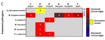
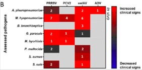
Figures 3 and 4 - Interactions between pathogens and the impact on clinical signs observed (Source: Saade et al., 2020)
Bacterial co-infections with M. hyopneumoniae
In relation to bacterial co-infections, the importance of M. hyopneumoniae is as crucial as with viruses:
- Actinobacillus pleuropneumoniae (App): in pigs simultaneously infected with M. hyopneumoniae and App, clinical signs and lung lesions are severe and correspond to the pathogenicity of the two bacterial strains combined. However, if the pigs are first infected with M. hyopneumoniae and later with App, these will be particularly affected with very severe clinical signs (Marois et al., 2009). Reducing the impact of M. hyopneumoniae through effective vaccination has been shown to also reduce the impact of pleuropneumonia caused by App, reflected in both reduced pleurisy and severity of lesions (Velazquez and Gale, 2019), demonstrating the importance of the interaction between the two pathogens.
- Pasteurella multocida: one of the major receptors on epithelial cells for this pathogen is a sugar (L-fucose). In healthy lungs there is no L-fucose expressed. However, if infection with M. hyopneumoniae occurs on the ciliated epithelium in the lungs, the sugars are modified, meaning that P. multocida has access to L-fucose receptors and therefore results in increased adhesion (Park et al., 2016).
In general, M. hyopneumoniae has a large role in increasing the severity of respiratory disease on farms where other pathogens are also present. Other bacteria such as Bordetella bronchiseptica, Haemophilus parasuis, Trueperella pyogenes, Streptococci or Staphylococci are also commonly found in field outbreaks of EP (Maes et al., 2018).
What is the economic impact of enzootic pneumonia in pigs?
M. hyopneumoniae is known to be one of the most prevalent swine pathogens worldwide, causing substantial economic losses within the swine industry (Maes et al., 2018). Enzootic pneumonia has a large economic impact on the pig industry, primarily due to the cost of treatment, reduced performance and increased mortality due to secondary infections (Holst et al., 2015).
The economic impact primarily derives from the decreased performance in production parameters, including:
- Reduced average daily gain (ADG)
- Reduced feed conversion ratio (FCR)
- Increased mortality
- Increased use of antibiotics to control EP and other pathogens involved in the PRDC (Maes et al., 2008)
Straw et al. (1989) demonstrated that pneumonia due to M. hyopneumoniae caused a decrease in ADG and feed efficiency by 17% and 14% respectively. This is reflected in increased days to slaughter, therefore increasing cost of production.
Calculating the economic impact due to M. hyopneumoniae specifically is difficult to estimate due to the contribution of numerous co-infections (Maes et al., 2018). The cost has been shown to be higher if other respiratory pathogens such as PRRS or SIV are also present along with M. hyopneumoniae (Haden et al., 2012). A recent study has demonstrated that the severity of the impact on productive performance, and therefore economic losses, is correlated with the percentage of lung consolidation caused by M. hyopneumoniae, reporting a negative correlation between lung consolidation lesions and ADG, where a 1% increase in lesion area corresponded to a decrease of 1.8 g (Ferraz et al., 2020). Therefore, these few studies are similar in the economic impact of M. hyopneumoniae noted; however, it is important to remember that this value will always depend on the extent and severity of lesions.
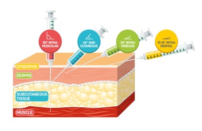
Correct angles of vaccination
The role of vaccination programmes in controlling enzootic pneumonia in swine
Vaccination is commonly used at weaning to control EP and has been shown to improve the daily weight gain by 2–8% and the feed conversion ratio by 2–5%, therefore reducing losses due to EP (Segales et al., 2008). There are numerous vaccines available which vary in the vaccination schedule, but the ultimate goal should be reduction of lung lesions and performance losses due to M. hyopneumoniae.
Vaccination with an efficient vaccine has been shown to be effective at reducing both the percentage of pigs with EP-like lesions and the severity of these lesions, as demonstrated by the Madec index which is calculated using a modified Madec score. The Ceva Lung Program (CLP) is a tool for analysis of the lungs in the slaughterhouse and has been used in many swine-producing countries to demonstrate these reductions.
Vaccination is also associated with reduced use of antimicrobials to control disease (Maes et al., 2008), contributing to achieving targets in reducing antimicrobial use across the industry. Vaccination combined with good management practices will help to reduce the impact of the pathogen as well as co-infections on farms, working to reduce the lung lesion prevalence and severity, which has been shown to be correlated with production losses.
Key Takeaways
- Co-infections can increase disease severity: Mycoplasma hyopneumoniae modifies the immune response and facilitates co-infections with viruses such as PRRSV, SIV and PCV2, as well as bacterial pathogens worsening clinical signs and lung lesions.
- Economic losses stem from reduced performance: One study reported reductions of 17% in average daily gain (ADG) and 14% in feed efficiency, resulting in more days to slaughter and higher production costs.
- Lung lesion severity correlates with production losses: A 1% increase in lung consolidation area corresponds to a 1.8 g decrease in daily gain, making slaughterhouse lung scoring a valuable monitoring tool.
- All-in-all-out management reduces pathogen pressure: Practising all-in-all-out systems, controlling stocking density and avoiding introduction of infected animals are integral to an effective enzootic pneumonia control plan.
- Vaccination improves performance and reduces antimicrobial use: Vaccination at weaning has been shown to improve daily weight gain by 2–8% and feed conversion ratio by 2–5%, while is associated with reduced antimicrobial use.
